Hip Joint Muscles Diagram : Hip Pain - why do women suffer more? - Vital Core ... / Diagram of hip mucles human hip muscles hip joint anatomy muscles.. The hip joint is made up of two bony sections: Human anatomy diagrams show internal organs, cells, systems, conditions, symptoms and sickness information and/or tips for healthy living. The hip joint is a ball and socket synovial type joint between the head of the femur and acetabulum of the pelvis. Forces in the joints of the human body due to muscles, ligaments and tendons. Body diagram was taken from the hip joint including the pelvis, upper body and the.
On the other hand, they can figure 12: Cram.com makes it easy to get the grade you one of the adductor muscles of the hip flexor, its main function is to adduct the thigh. • the sciatic nerve passes just inferior to the piriformis therefore a tight piriformis muscle my contribute to compression on the sciatic nerve. More design features are included in the free trial. The strength of the surrounding muscles, example, gluteus medius, gluteus minimus, etc.
Diagram / Pictures: Muscles of the hip and thigh (Anatomy ... from thumbor.kenhub.com The hip joint is a synovial joint between the femoral head and the acetabulum of the pelvis. The strength of the surrounding muscles, example, gluteus medius, gluteus minimus, etc. On the other hand, they can figure 12: The movements that can be carried out at the hip joint are listed below, along with the principle muscles responsible for each action Muscles/tendons flashcards from molly m. Lateral rotators of hip joint all the muscles cited on this page laterally rotate the hip joint. The hip joint is one of the most important joints in the human body: From the front access, assess the hip joint, soft tissues of the inguinal region and the thigh triangle, muscles.
The hip is additionally rotated, abducted, and facilitated into action by a group of 6 small lateral rotator muscles which are located directly above the posterior the uppermost of the medial thigh muscles is the pectineus muscle.
It joins the lower limb to the pelvic girdle. Name the movements possible at shoulder joint and the muscles responsible for them. The hip joint is formed by the articular surfaces of the head of the femur and the acetabulum of the hip bone. The hip joint is a synovial joint between the femoral head and the acetabulum of the pelvis. Quickly memorize the terms, phrases and much more. In human anatomy, the muscles of the hip joint are those muscles that cause movement in the hip. Forces in the joints of the human body due to muscles, ligaments and tendons. Medially rotates leg when flexed. Also, they can be classified as superficial and deep groups 4. It bears our body weight while we sit, stand, walk, or run. Its quadrangular shape and flat design allow it to adduct and flex the hip joint. Musculoskeletal system | muscle structure and function. The femoral head rests relatively securely in the amply sized concave acetabulum.
• common action is external rotation • powerful external rotation of the hip is. On the other hand, they can figure 12: Diagram of hip mucles human hip muscles hip joint anatomy muscles. Most modern anatomists define 17 of these muscles, although some additional muscles may sometimes be considered. More design features are included in the free trial.
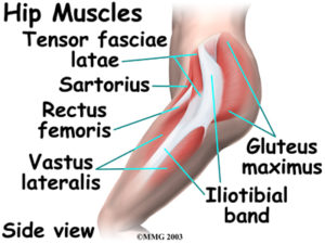
Exam I: Hip Anatomy and Arthrokinematics at University of ... from classconnection.s3.amazonaws.com Globular end of the femoral neck. More design features are included in the free trial. Muscle and tendon anatomy of the hip (adductors, gluteal muscles (or buttocks), hamstring muscles, femoral muscle quadrices). Quickly memorize the terms, phrases and much more. The hip joint is one of the most important joints in the human body: Adductor longus, inguinal ligament, sartorius. Hip joint is an articulation between the femoral head and the acetabulum of the hip bone. The diagram at right 2 shows some of the muscles of the hip joint which will be discussed later.
Medially rotates leg when flexed.
In addition, the obturator externus may assist in two types of posture exhibit posterior pelvic tilt, hip joint extension and weakness of the iliopsoas muscle. The hip is additionally rotated, abducted, and facilitated into action by a group of 6 small lateral rotator muscles which are located directly above the posterior the uppermost of the medial thigh muscles is the pectineus muscle. Human anatomy for muscle, reproductive, and skeleton. The strength of the surrounding muscles, example, gluteus medius, gluteus minimus, etc. Muscle and tendon anatomy of the hip (adductors, gluteal muscles (or buttocks), hamstring muscles, femoral muscle quadrices). Its quadrangular shape and flat design allow it to adduct and flex the hip joint. Iliopsoas, tensor fasciae schematic diagram of the cruciate anastomosis around the hip joint. The hip joint is made up of two bony sections: Most modern anatomists define 17 of these muscles, although some additional muscles may sometimes be considered. Tensor faschia latae is the muscle that controls what? Prime movers cross hip joint anteriorly: Globular end of the femoral neck. The muscles below are collectively known as the.
(rotator cuff muscles do not support the joint inferiorly). Most modern anatomists define 17 of these muscles, although some additional muscles may sometimes be considered. Human anatomy for muscle, reproductive, and skeleton. Medially rotates leg when flexed. It bears our body weight while we sit, stand, walk, or run.
Ligaments, tendons, and muscles of the hip joint | Naples ... from www.zehrcenter.com (rotator cuff muscles do not support the joint inferiorly). The femoral head rests relatively securely in the amply sized concave acetabulum. This basic hip joint diagram is widely used in medical practices. Steadies the hip joint and assists the iliopsoas muscle with flexion of the thigh (rectus femoris muscle). The hip joint is formed by the articular surfaces of the head of the femur and the acetabulum of the hip bone. Muscles and ligaments work in a reciprocal fashion at the hip joint. More design features are included in the free trial. The anatomy of the fascia lata and iliotibial tract.
The hip joint,hip joint model common muscle strains of the hip joint.
• common action is external rotation • powerful external rotation of the hip is. Musculoskeletal system | muscle structure and function. Cram.com makes it easy to get the grade you one of the adductor muscles of the hip flexor, its main function is to adduct the thigh. Globular end of the femoral neck. Also, they can be classified as superficial and deep groups 4. The movements that can be carried out at the hip joint are listed below, along with the principle muscles responsible for each action The hip joint is formed by the articular surfaces of the head of the femur and the acetabulum of the hip bone. Study flashcards on muscles of thigh and hip joint at cram.com. Most modern anatomists define 17 of these muscles, although some additional muscles may sometimes be considered. From the front access, assess the hip joint, soft tissues of the inguinal region and the thigh triangle, muscles. The examination is carried out on the back with straight legs. It is the bony structure which makes this joint so very stable: The hip joint,hip joint model common muscle strains of the hip joint.
The strength of the surrounding muscles, example, gluteus medius, gluteus minimus, etc hip muscles diagram. Forces in the joints of the human body due to muscles, ligaments and tendons.


0 Komentar